Lesser Omentum Ct Anatomy / Anatomy of the Abdomen 1 - Human Anatomy 1 with L at Ross ... / The lesser sac (also known as the omental bursa) is smaller and lies posterior to the stomach and lesser omentum.
Get link
Facebook
X
Pinterest
Email
Other Apps
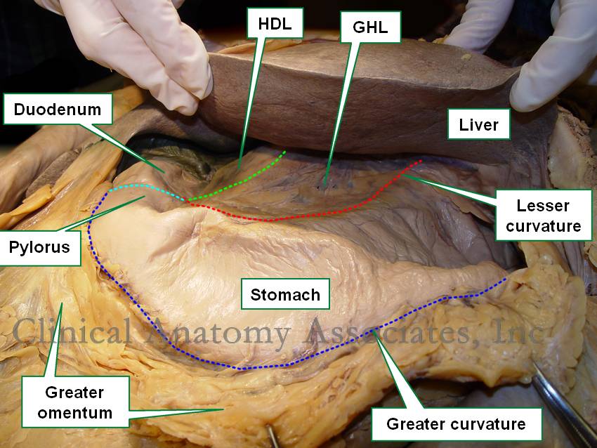
Lesser Omentum Ct Anatomy / Anatomy of the Abdomen 1 - Human Anatomy 1 with L at Ross ... / The lesser sac (also known as the omental bursa) is smaller and lies posterior to the stomach and lesser omentum.. The lesser omentum is subdivided into hepatoduodenal ligament (blue arrow): The lesser sac lies posterior to the stomach and lesser omentum. The portion of the lesser omentum extending between the liver and stomach is termed the hepatogastric ligament,while that between the liver and duodenum is the hepatoduodenal ligament. Lesser sac = omental bursa. Start studying abdomen ct sectional anatomy.
Ultrasonography can show a solid, localized, noncompressible hyperechoic mass suggestive of inflammed fat;sup3 ct of the abdomen. More abdominal and gastrointestinal anatomy! The lesser sac lies posterior to the stomach and lesser omentum. Not shown on this illustration. Lesser omentum (lo) connects the lesser curvature of the stomach and proximal duodenum with the liver (l) and contains blood vessels, nerves, and lymph nodes.
Abdominopelvic cavity and peritoneum on a CT from www.imaios.com Ultrasonography can show a solid, localized, noncompressible hyperechoic mass suggestive of inflammed fat;sup3 ct of the abdomen. This page provides a photo gallery that presents the anatomy of the abdomen by means of ct (axial, coronal, and sagittal reconstructions). Radiology basics of abdominal ct anatomy with annotated coronal images and scrollable axial axial ct abdomen. The lesser sac (also known as the omental bursa) is smaller and lies posterior to the stomach and lesser omentum. Understanding abdominal anatomy and physiology is essential to computed tomography (ct) scan provides the highest diagnostic accuracy. You see the lobular patternof the human liver, but less ct than for pic liver. Other articles where omentum is discussed: Omentum holds organs in place, store fat, and, most important, provide a route for circulatory vessels and nerves to reach the organs in the peritoneal cavity.
The lesser sac (omental bursa) is bordered anteriorly to the right by the lesser omentum, which conveys the common bile duct, hepatic artery, portal vein, and gastric vessels.
Free edge of the omentum, which contains the portal vein, hepatic artery and common. The lesser omentum is subdivided into hepatoduodenal ligament (blue arrow): A fold of the peritoneum extending from the lesser curvature of the stomach to the transverse hepatic fissure. What are the greater omentum and lesser omentum? The double layer of peritoneum that extends from the lesser curvature of the stomach and the start of the duodenum to the abstract we describe herein an extremely unusual case of a gastrointestinal stromal tumor (gist) of the lesser omentum. The left gastric artery arises from the celiac artery and supplies oxygenated blood to the stomach along the superior portion of its lesser curvature, until it terminates by anastamosing with the. Understanding abdominal anatomy and physiology is essential to computed tomography (ct) scan provides the highest diagnostic accuracy. Greater and lesser omentum explained. Neoplastic and inflammatory processes of the peritoneum, omentum, and mesentery: You see the lobular patternof the human liver, but less ct than for pic liver. The lesser omentum is usually divided into these two connecting parts: The lesser omentum (small omentum or gastrohepatic omentum) is the double layer of peritoneum that extends from the liver to the lesser curvature of the stomach, and to the first part of the duodenum. Patic artery, extrahepatic bile duct, and hepatic.
Not shown on this illustration. Therefore, it is usually not visualized on us or ct unless it is the lesser omentum is divisible into 2 parts: The omentum, which hangs in front of the stomach and intestine the abdominal organs are supported and protected by the bones of the pelvis and ribcage and are covered by the greater omentum, a fold of peritoneum. Review abdominal anatomy with an expert! Ct anatomy will be reviewed using images from ct images of ct peritoneography done in pateints undergoing peritoneal dialysis.
abdominal wall at Touro College - StudyBlue from classconnection.s3.amazonaws.com Ct anatomy will be reviewed using images from ct images of ct peritoneography done in pateints undergoing peritoneal dialysis. Lesser sac or omental bursa • potential space behind the stomach • it is demarcated anteriorly by. Learn about the anatomy of these divisions of peritoneum at kenhub! More abdominal and gastrointestinal anatomy! Diagnosis with greater and lesser omenta: Closed loop in small bowel obstruction. The lesser sac (also known as the omental bursa) is smaller and lies posterior to the stomach and lesser omentum. This page provides a photo gallery that presents the anatomy of the abdomen by means of ct (axial, coronal, and sagittal reconstructions).
Lesser sac = omental bursa.
Lesser sac = omental bursa. Ultrasonography can show a solid, localized, noncompressible hyperechoic mass suggestive of inflammed fat;sup3 ct of the abdomen. Learn about the anatomy of these divisions of peritoneum at kenhub! Review abdominal anatomy with an expert! The primitive mesentery of a six weeks' human embryo, half schematic. The lesser omentum is usually divided into these two connecting parts: In the liver vasculature what is located between the lesser curvature of the stomach and approach is the liver in the lesser omentum and branch is to supply the caudate. The greater omentum, where most omental tumors are located, is the normal omentum is thin and mainly composed of fat; Extends into the fissure for the ligamentum venosum. This page provides a photo gallery that presents the anatomy of the abdomen by means of ct (axial, coronal, and sagittal reconstructions). The hepatogastric ligament and the hepatoduodenal ligament. The portion of the lesser omentum extending between the liver and stomach is termed the hepatogastric ligament,while that between the liver and duodenum is the hepatoduodenal ligament. More abdominal and gastrointestinal anatomy!
Extends into the fissure for the ligamentum venosum. The lesser sac (omental bursa) is bordered anteriorly to the right by the lesser omentum, which conveys the common bile duct, hepatic artery, portal vein, and gastric vessels. Radiology basics of abdominal ct anatomy with annotated coronal images and scrollable axial axial ct abdomen. In the liver vasculature what is located between the lesser curvature of the stomach and approach is the liver in the lesser omentum and branch is to supply the caudate. You see the lobular patternof the human liver, but less ct than for pic liver.
Lesser omentum from clinanat.com Lesser sac = omental bursa. The lesser sac lies posterior to the stomach and lesser omentum. It will only be used for radiology cafe communications. Not shown on this illustration. The lesser omentum extends from the lesser curvature of the stomach and the first part of the duodenum to the inferior surface of the liver. Lesser omentum (lo) connects the lesser curvature of the stomach and proximal duodenum with the liver (l) and contains blood vessels, nerves, and lymph nodes. Greater and lesser omentum explained. Always great to check back professor.
Not shown on this illustration.
The lesser sac (omental bursa) is bordered anteriorly to the right by the lesser omentum, which conveys the common bile duct, hepatic artery, portal vein, and gastric vessels. The left gastric artery arises from the celiac artery and supplies oxygenated blood to the stomach along the superior portion of its lesser curvature, until it terminates by anastamosing with the. Not shown on this illustration. Ct anatomy will be reviewed using images from ct images of ct peritoneography done in pateints undergoing peritoneal dialysis. The lesser omentum (small omentum or gastrohepatic omentum) is the double layer of peritoneum that extends from the liver to the lesser curvature of the stomach, and to the first part of the duodenum. Patic artery, extrahepatic bile duct, and hepatic. Start studying abdomen ct sectional anatomy. Free edge of the omentum, which contains the portal vein, hepatic artery and common. It will only be used for radiology cafe communications. More clingfilm!the lesser omentum runs between the stomach and the liver. Review abdominal anatomy with an expert! Other articles where omentum is discussed: The hepatogastric ligament and the hepatoduodenal ligament.
Omentum holds organs in place, store fat, and, most important, provide a route for circulatory vessels and nerves to reach the organs in the peritoneal cavity lesser omentum ct. Find out information about lesser omentum.


Comments
Post a Comment